Getting a Closer Look Inside Animals: Computed Tomography Comes to the Detroit Zoological Society

As zoo veterinarians, we recognize the importance of identifying animals with health problems as early as possible. Fortunately, the Detroit Zoological Society has exceptional zookeepers who attentively look after each animal in their care and alert the veterinary team whenever they suspect there may be a problem. While subtle changes in demeanor, appetite, fecal and urinary output, and activity level can be key indicators of illness in an animal, most of our patients are very good at hiding their symptoms. In order to get a more comprehensive understanding of an animal’s health, we often rely on diagnostic tests, such as physical examination, bloodwork and cultures for bacteria.

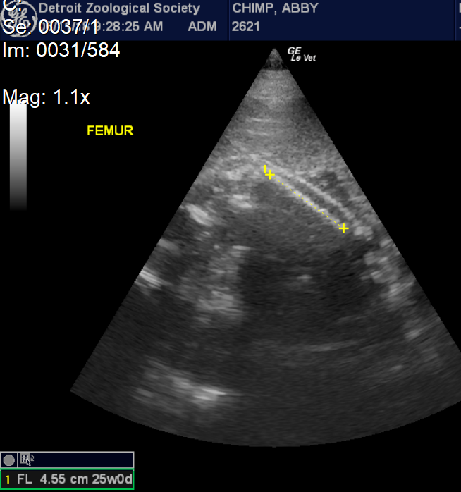
When the Ruth Roby Glancy Animal Health Complex opened in 2004, the radiology suite was equipped with a state-of-the-art radiology unit designed for use in human hospitals.  With this upgrade, we found that we increasingly relied on diagnostic imaging (radiographs and ultrasound) to make diagnoses and shape our treatment plans.  In fact, we take x-rays during almost every diagnostic examination, on patients as small as dart frogs and as large as bison.

Since the early 2000s, imaging technology has been rapidly advancing, and by upgrading equipment and adding new technologies, the Detroit Zoological Society has stayed on the cutting edge of veterinary care.  This includes having ultrasound probes designed for patients of all shapes and sizes, digital dental radiography and portable x-ray equipment that can go out into the Zoo to image animals who are difficult to move to the hospital. Despite these advancements, we still found it necessary to take patients to off-site facilities at least a few times each year for computed tomography (CT) or magnetic resonance imaging (MRI).

In late 2019, two very exciting things happened: first, a generous donor named Thomas A. Mackey came forward with an interest in funding a project that would have an immediate impact on animal care and welfare, and, secondly, we became aware of a revolutionary new computed tomography (CT) technology that had been developed in Ann Arbor.  One of the most important features of the new CT technology is that it is portable, and much more affordable and user-friendly than a full-sized CT system. Since our hospital was already equipped with the features and space necessary to install the new system, within just a few months, we were able to bring this exciting new technology to the Zoo.

The new Xoran Portable CT has been in use for only a few months, but it has already had a tremendous impact on patient care at the Detroit Zoo.  Adding CT to our diagnostic toolbox has increased the level of care that we can provide to animals at DZS exponentially. CT works by aiming a narrow beam of x-rays at a patient, while quickly rotating around them. The CT’s computer generates cross-sectional images, or “slices” of the body.  The images contain more detailed information than conventional x-rays.  Once the slices are generated, they can be digitally “stacked” together to form a 3-D image that allows for easier identification and location of basic structures as well as possible tumors or abnormalities.

Here are just a few examples of how this technology is helping us give animals the best possible care:

CASE #1
CT imaging is especially well suited for visualizing the teeth and bones of the jaw. A male aardvark was due for a routine checkup. He had been eating fine, and there was no reason to suspect that he had dental disease. However, aardvarks often have problems with their teeth, so we decided to use the CT machine to scan his head. The images collected showed that he had areas of bone breakdown around the roots of three separate teeth. Treatment was able to be provided before his condition progressed to a point where he was showing signs of discomfort.

Picture1    2

CASE #2
This adult McCord’s box turtle was imaged during a routine examination. The shell covering the body can make radiographs hard to interpret, but CT imaging allows us to see inside of the turtle.

McCord’s box turtle: a. Image of the head and forearms, b. image from the side showing the head and neck folded into the shell, c. 3D reconstruction of the face and front limbs seen in image a.

a. 1  b. 23                                                 c.

CASE #3
CT imaging has also proved helpful for several avian patients. One of the cinereous vultures living at the Zoo had a mass (red star) growing on the toe pictured below. The mass needed to be removed, but in order to plan for surgery, we needed to understand if the mass was superficial or more invasive and involved the soft tissues and bone beneath. CT imaging provided better detail for seeing small changes in the muscles and ligaments surrounding the mass. After evaluating the images, we were able to plan a surgical approach to remove the mass, and any adjacent tissue of concern. The vulture is doing great post-operatively and already back in his home!

Screen Shot 2020-02-12 at 10.25.27 AM    Screen Shot 2020-02-12 at 10.25.37 AM

CASE #4
We currently have four red pandas living at the Detroit Zoo. The oldest is a 14-year-old female named Ta-Shi. During her recent routine examination, we noticed that one of her large molar teeth appeared darker than normal and was cracked on the surface. Within a few moments, we were set up and ready to collect CT images of her head and teeth. The images showed that the tooth was infected at the root, a problem that was likely causing discomfort. The tooth was also broken, meaning it needed to be removed in several pieces. After the tooth was extracted, a repeat CT showed us conclusively that all of the roots had been completely removed.

1    2

We are extraordinarily grateful to have state-of-the-art equipment at hand to care for animals at the Detroit Zoo. The recent financial gift that made the addition of CT possible has improved our ability to see small changes more clearly, detect problems earlier and fine-tune treatments. With this tool, we will continue to ensure that animals live long, healthy lives and thrive within our care. We cannot say thank you enough to Thomas A. Mackey for his incredibly generous donation!

– Dr. Ann Duncan is the director of animal health for the Detroit Zoological Society and oversees the Ruth Roby Glancy Animal Health Complex.


Veterinary Care: Repairing a Warthog’s Fractured Tusk

The most distinctive features of warthogs are the gently curving tusks that protrude from either side of their face. Each has two pairs of tusks, which are actually constantly growing canine teeth, with the upper pair usually much longer than the lower.

During a recent routine examination of a 3-year-old female warthog named Sansa (yes, after Game of Thrones) by veterinary staff at the Detroit Zoological Society’s Ruth Roby Glancy Animal Health Complex, we noticed that she had fractured one of her lower tusks below the gumline. We couldn’t see the tooth, so we used a metal dental instrument to locate the end of the tooth and then took a radiograph. We could tell that the fracture had occurred very recently and were concerned that the open end of the tooth might allow bacteria to enter. If an infection reaches the base of the tooth, extraction may become necessary, and we wanted to do whatever we could to avoid this.

The size and shape of a warthog’s tusk is similar to the canine tooth of a domestic horse, and we knew that our equine dentist frequently treats his patients for broken canines. We asked Dr. Tom Johnson to come to the Detroit Zoo to help us repair her tooth. Her follow-up exam was less than three weeks since her first exam and already the tooth had grown enough that it was visible at the gumline, which made treatment much easier than expected. We started by cleaning the surface of the tooth and could see that the opening was very small, making infection less likely to occur.

The procedure was very much like having a cavity filled: Dr. Johnson used a dental drill to cup out the area around the opening and then used dental materials to seal and fill the tooth. The filling will remain protected within this recessed area while the tooth continues to grow and be used. The radiographs show that there is very good blood supply to the tooth and we expect that Sansa will be able to heal completely now that the tooth has been repaired. To be certain, we will examine her again in a year, and will take another radiograph to check for any concerns.

– Dr. Ann Duncan is the director of animal health for the Detroit Zoological Society and oversees the Ruth Roby Glancy Animal Health Complex.

Veterinary Care: Baby Jane’s Prenatal Check-ups

While newborn photos of a female baby chimpanzee have gone viral on our social media accounts, they weren’t the first images taken of little Jane. During mom Abby’s 33-week pregnancy, Detroit Zoological Society staff performed eight ultrasounds of the baby, who is named after legendary primatologist and anthropologist Dr. Jane Goodall.

Staff works diligently with the great apes who live at the Detroit Zoo to develop behaviors that allow us to monitor their health. The gorillas and chimpanzees open their mouths to let us look at their teeth, show us their hands and feet and lean against the mesh to allow the administration of vaccines. Most of the chimpanzees will press their chests toward the mesh so we can take images of their hearts with an ultrasound probe.  Abby quickly learned to position herself and allow us to put the probe on her belly so that we could monitor her growing fetus. After a few practice sessions, we invited an OB (obstetrical) ultrasound technician to the Zoo to take the standard measurements collected during pregnancy in human women.

Abby was a cooperative patient and always appeared excited to see us. She would prop herself on a ledge and eat peanuts during each exam, allowing the peanut shells to pile up on her growing belly.  There are limits to the ways we can position the probe, and we were not always able to get every measurement at every visit. In the early months, we were able to measure the length of the fetus from the crown to the rump; as the baby grew, we measured the circumference of the head and abdomen and length of the long bones, including the femur and humerus. We were also able to see the position of the fetus and measure the heart rate. With each exam, we added data to our growth charts, and were pleased to see steady growth and development. We also became increasingly confident that the baby was a girl.

Abby is the third chimpanzee mom that has allowed us to conduct obstetrical ultrasounds, and since 2008 we have been able to collect measurements from three pregnancies, including youngsters Ajua and Akira. Using these measurements and data from two scientific publications, we were able to make a solid prediction of Abby’s due date – July 14, the date of the first annual World Chimpanzee Day! As this date approached, animal care staff began round-the-clock checks to look for signs of labor. Just three days before the due date, we performed a final ultrasound exam. We were pleased to see that the baby was still growing according to expectations. We could see her face and watch her open and close her mouth and wiggle her arms and legs. Most importantly, we could see that the baby had a strong heartbeat and was positioned with her head down, which is the correct position for a normal delivery.

Anyone who has anticipated the delivery of a baby knows that due dates are not an exact science. But Abby delivered her baby at 12:01 a.m. on July 14, one minute into the day predicted as her due date, and the delivery was without complication. Being able to monitor babies during pregnancy allows us to prepare for any issues that might arise, and to intervene if needed. Abby is a wonderful mom, and is taking good care of Jane. She seemed excited to show off her new baby to the other chimpanzees, and held her against the window for everyone to see. We look forward to watching her grow and thrive in her habitat at the Great Apes of Harambee.

– Dr. Ann Duncan is the director of animal health for the Detroit Zoological Society and oversees the Ruth Roby Glancy Animal Health Complex.

Preventative Care Ensures Quality of Life for Zoo Animals

Spring has sprung! This time of year brings April showers, blooming daffodils, lawns to mow and the delightful warmth of the sun finally reaching our skin. It also brings mosquitos, an aspect of the season I do not enjoy. While their numbers are still low, they are staging a comeback and before long, I’ll be carrying bug spray with me in my bag.

With mosquitos comes heartworm season, a time when veterinarians and pet owners are reminded of the importance of the preventive care they provide the animals in their lives. Taking preventive measures is the best way to avoid bigger, more difficult problems in the future. At the Detroit Zoo, we have an extensive preventive medicine program that addresses vaccinations, parasite prevention, nutrition and regular examinations.

Vaccinations are one of the most important tools available to prevent disease in animals and people. In zoo medicine, we refer to literature to determine the safest and most effective vaccines available for the species in our care. We also communicate frequently with zoo and animal health colleagues to better understand diseases of concern. Each month, we participate in a One-Health conference call with veterinarians from the Michigan Department of Agriculture and Rural Development, Department of Natural Resources, U.S. Department of Agriculture’s Animal Plant Health Inspection Service (APHIS) and public health officials from the Department of Health and Human Services. We discuss the diseases currently posing a risk to humans and animals alike, talk about the most common diseases and discuss newer threats, such as avian influenza and Lyme’s disease. At the Detroit Zoo, we use vaccines to prevent many of these diseases, including rabies, distemper, leptospirosis and tetanus. Contagious diseases can be introduced to the animals by wild raccoons, skunks, rodents and feral and pet cats that enter the Zoo, and it’s important that we protect susceptible zoo animals. We use vaccines developed for domestic horses, cows and cats to protect the zebras, elands and lions.

Heartworm disease is another focus of preventive care. Heartworm is transmitted by mosquitos and can go undetected until infection poses a serious health risk. You may know that heartworm disease infects dogs and cats, but you may be surprised to learn the array of animals at the Zoo that can be affected. Seals, arctic foxes, bush dogs, lions, tigers, otters and wolverines are all treated year-round with a monthly preventive treatment. I am happy to report that in my more than 20-year career, we have never had an animal test positive for heartworm disease at the Zoo. If your dog or cat is not being treated for heartworm, it’s important to have them tested first, and then to start them on prevention as soon as possible. It’s never too late to get started.

Examinations are another important part of preventive care. Every day, the animal care staff observes each animal at the Zoo, noting any abnormal appearance or behavior to the veterinary staff. Many health problems are detected through these diligent observations, but we also periodically perform a  hands-on exam to be sure we aren’t missing anything. During examinations, we take radiographs to check for arthritis; feel for bumps and skin problems; examine the eyes with an ophthalmoscope, and use ultrasound to check the heart and abdominal organs. We do a careful dental exam and scale and polish the teeth – this is often the most important thing we do to improve health. During these exams, we also collect a blood sample and run tests to get an impression of overall health and check for diseases of concern.

One of the most rewarding things as a veterinarian is to find a problem before it becomes life threatening and fix it. With close observations, exams and regular care, we tip the odds in favor of finding problems early and being able to intervene, but this is not always possible. Last year, I lost my beloved golden retriever to cancer after a very sudden illness. Even though I miss her, I am at peace because I know that there isn’t anything I could have done to change her end-of-life story. Here at the Zoo, the animal care and veterinary teams strive to do the same. We do all that we can to find problems early, to treat illness when possible, and to help animals nearing the end of their lives maintain the best-possible quality of life. I’m proud of the care that we provide the animals at the Zoo, and glad to share these stories.

– Ann Duncan is the director of animal health for the Detroit Zoological Society and oversees the Ruth Roby Glancy Animal Health Complex.

Veterinary Care: Cutting-edge Technologies in Amphibian Conservation

Last week I was able to attend a very exciting advanced conservation training course to learn assisted reproductive technologies (ART) in amphibians. The meeting was hosted by the Omaha Zoo, and was offered by the Amphibian Taxon Advisory Group (ATAG). I was one of a small group of zoo and conservation scientists invited to learn cutting-edge technologies that have the potential to have a tremendous impact on the conservation of endangered amphibians.

At the Detroit Zoo, we’ve been using hormone treatments to help with reproduction in Wyoming toads and Puerto Rican crested toads for more than 10 years. Recently, Dr. Andy Kouba from Mississippi State University has been able to modify these treatments for use in other species, and to develop techniques for collecting eggs and sperm for in-vitro fertilization (IVF). Two years ago, I traveled to his laboratory to learn these techniques for dusky gopher frogs, and within a few months we became the third zoo to reproduce amphibians via IVF.

The goal of the ART course was to provide in-depth information concerning reproduction in frogs, toads and salamanders. This course was taught by Dr. Kouba and two of his colleagues, and represented the first time that husbandry staff, veterinarians and conservation researchers have come together to discuss assisted reproductive technologies. The attendees brought together a wealth of knowledge, and experiences working with hellbender salamanders, Chinese giant salamanders, Oregon-spotted frogs and others. We talked about the impacts of hibernation, temperature, humidity and social cues on reproduction, and the challenges we have encountered in the past. We learned about the historic use of hormones in amphibians, and how this information can be adapted to new species. We also learned how to gently collect spermic urine from male amphibians, and to stimulate females to lay eggs. We learned how to examine developing tadpoles under a microscope, and cyroperservation techniques for sperm. We also learned how to use ultrasound to monitor egg development in females. During the week, we put these skills to practice, and were able to produce fertilized eggs from Puerto Rican crested toads, American toads, tiger salamanders and Asian black spiny toads (for the first time!).

At the Detroit Zoo, we have number of very endangered species of amphibians, including some that have never or only rarely reproduced outside of the wild. Over the next few months, we will be able to start using cryopreservation to save genetics from these critically endangered species. The skills learned at the ART course will allow us to continue to be leaders in amphibian conservation, and to hopefully successfully breed Japanese giant salamanders, giant waxy tree frogs, and other endangered species at the National Amphibian Conservation Center.

– Dr. Ann Duncan is the director of animal health for the Detroit Zoological Society and oversees the Ruth Roby Glancy Animal Health Complex.

Veterinary Care: My, What Big Teeth You Have!

Dental care is a very important component of maintaining the health and welfare of the animals at the Detroit Zoo. Many of the animals in our care live longer than they would in the wild, and we need to ensure that they maintain healthy, strong teeth throughout their lifespans.

Some of our patients are cooperative; for example, the polar bears and chimpanzees have been trained to open their mouths wide for a quick visual exam, and the seals allow us to brush their teeth daily to reduce tartar build up. But it’s not always that easy – our patients all have very different mouths, and some teeth are easier to see than others. During every health check, we make sure to do a thorough dental exam.


Most of our carnivores and reptiles have mouths that open wide and teeth that are easy to examine. Aardvarks, kangaroos and pigs are examples of animals that have elongated, deep mouths that are very hard to visualize, but we use a variety of tools to get the job done. A laryngoscope with a very long blade or small endoscope can be used to see in the dark, tight spaces inside the mouth, such as with a Meishan pig. Pigs can develop dental problems as they age, so it’s important to check each tooth and treat any potential problems early.

Our patients also have different kinds and shapes of teeth. The teeth of carnivores and herbivores are shaped differently, and their structure can vary as well. We look up the dental formula (how many incisors, premolars and molars a species has) and anatomy of the teeth before exams, and we have developed a new dental exam form to help us identify the teeth and record any problems we find. For example, beavers have front incisors that grow continuously, and we check their teeth frequently for overgrowth.

Some of our patients have very, very large teeth, like Kisa, a 13-year-old female Amur tiger. During Kisa’s most recent health check, we were able to see that there was swelling around her lower left canine tooth. We scheduled an exam with our visiting veterinary dentist, Dr. Ben Colmery. He made a small incision along the side of the tooth and found an area where the enamel had eroded away. This type of enamel resorption is common in older domestic cats, and has been seen in our geriatric zoo patients as well. If left untreated, serious problems can develop that can make it necessary to extract the tooth. Dr. Colmery smoothed the lesion and applied a special paste to help stimulate healing. We also scaled and polished all of her teeth to remove tartar and plaque.

Our goal in the animal health department is provide the best possible care to all of the wonderful animals that live at the Detroit Zoo. We work to understand the kinds of health problems found in zoo animals, and to prevent these problems whenever possible. Finding dental disease and other health problems early allows us to be proactive in our efforts to maintain excellent health and welfare.

– Ann Duncan is the director of animal health for the Detroit Zoological Society and oversees the Ruth Roby Glancy Animal Health Complex at the Detroit Zoo.

Veterinary Care: Get Ready to Run Wild

This year marks the 20th anniversary of Run Wild for the Detroit Zoo, and we anticipate it will be bigger and better than ever. This is a fun, family-friendly fundraising event that encourages our friends and Members to enjoy a healthy dose of exercise – through either a 5K, 10K, Too Wild combo or Fun Walk. Run Wild is also a great way to support the health of the animals at the Zoo.

The race started as a unique collaboration between the Southeastern Michigan Veterinary Medical Association and the Detroit Zoological Society. During the first 10 years, proceeds from Run Wild were put toward a $1 million endowment dedicated to supporting the purchase of equipment for the Zoo’s Ruth Roby Glancy Animal Health Complex. Each year, the revenue from this endowment and proceeds from Run Wild are used to purchase and upgrade our diagnostic and treatment capabilities. This funding has allowed us to maintain cutting-edge health technologies in the Zoo. Over the years, we have purchased some very important equipment, including a two-headed teaching microscope, digital radiology, an ultrasound machine and a portable blood analyzer.

Last year we purchased a hand-held portable dental X-ray unit. Dental care is a very important component of our efforts to keep animals as healthy as possible. The new unit looks like a futuristic ray gun, and delivers a narrow beam of X-ray energy to a very small plate designed to be put inside the mouth of our patients. With this technology, we are able to take very detailed images of individual teeth or areas of concern, and to find dental problems early, before they become serious. The unit is small enough that it can easily be transported to our patients throughout the Zoo’s 125-acre grounds. We have used it to image teeth from wolverines to guanacos, lions and other animal patients of all shapes and sizes. We have even used it to take images of the bellies of frogs and toads!

Last year we also purchased a new patient monitoring tool called an EMMA (Emergency Mainstream Analyzer). This tiny piece of equipment provides us with very valuable information during anesthetic procedures. With each exhalation, the EMMA measures the amount of CO2 in the breath. Any change in body position or physiology that subtly compromises an animal’s ability to use oxygen is immediately known using this sensitive equipment. We use it for all of our procedures now, and find it invaluable.

I know this may all may sound very technical, but providing health care to the animals at the Zoo is a big job. The team at the Ruth Roby Glancy Animal Health Complex is very grateful to have the latest and greatest equipment to provide skilled and compassionate care. Join us for Run Wild, and know that you are supporting great care at your Zoo.

– Dr. Ann Duncan is the director of animal health for the Detroit Zoological Society and oversees the Ruth Roby Glancy Animal Health Complex.

Editor’s Note: New this year, all Run Wild participants will receive finisher medals! These medals feature a penguin to celebrate the Polk Penguin Conservation Center as well as an American flag ribbon to honor the anniversary of September 11. In addition to finisher medals, all participants will receive a reusable Detroit Zoo water bottle at the finish line. These bottles can be refilled at any of the free hydration stations on Zoo grounds, and there will be additional refilling stations available on race day. This is all part of our Green Journey to create a more sustainable future; last year we eliminated the sale of disposable water bottles at Detroit Zoo concessions, an effort that has kept 60,000 plastic bottles out of the waste stream.